Cholesteatoma is the disease for which I operate on the ear the most. It is a common cause of acquired deafness in children as well as adults. Sadly the condition is under-projected both by the paediatricians and the otolaryngologists. Also, there are hardly any symptoms to alert one about the disease in its early stages. Sometimes it is just a suspicion of a foul smell emanating from the ear or mild wetness or rarely a minimal hearing loss that prompts the patient to the doctor. More often they present in the late stages, with pain, giddiness, headache etc. which herald sinister life-threatening complications like meningitis or brain abscess. The condition is referred to as an unsafe ear in common parlance.
This disease is the finale of all those untreated nasal diseases that ultimately affect the ear. The eustachian tube connects the middle ear to the nasopharynx, the portion of the throat behind the nose. It helps equalise the air pressure in the middle ear with that of the atmospheric pressure and also drains out secretions, if any, in the middle ear, into the throat. Obstruction of the eustachian tube causes a spectrum of diseases in the ear according to the severity and duration.
Early and mild cases present with a blocked sensation of the ear. These patients have a shallow retraction of the eardrum due to the partial vacuum. Nasal decongestant medications are enough to cure this stage. The lining mucosa of the middle ear space may secrete mucoid secretions to neutralise the negative pressure. A minor surgery called ‘Myringotomy’ to remove the fluid is useful to cure the blocked sensation in the ear. In recurrent cases, ‘Grommets’ or ventilation tubes are inserted through the eardrum, in addition to treating the primary disease in the nose. Infection of this middle ear fluid may cause severe pain accompanied by fever, and may even result in the rupture of the eardrum and discharge of pus from the ear, if not treated by antibiotics and supportive management in time.
The ear canal is also lined by skin externally. Skin all over the body sheds its outer layers constantly. This desquamated skin, along with the wax, moves outwards in a natural mechanism. Long-standing negative pressure in the middle ear, if left unattended, can result in a retraction pocket in the eardrum and this hampers the drainage. Desquamated epithelium tends to collect in these pockets, and as this keratin increases in volume, the sac expands. Eventually, the retraction pocket meets the posterior bony wall of the middle ear, which it then erodes. Once the bone erosion starts, the disease takes a more sinister tone. The tiny bones responsible for the sound conduction- the malleus, the incus and the stapes, the balance organs of the ear, the facial nerve, brain, meninges etc., may all be at risk now. This condition is similar to what was described in my previous post – ‘Vexing wax and Keratosis’.
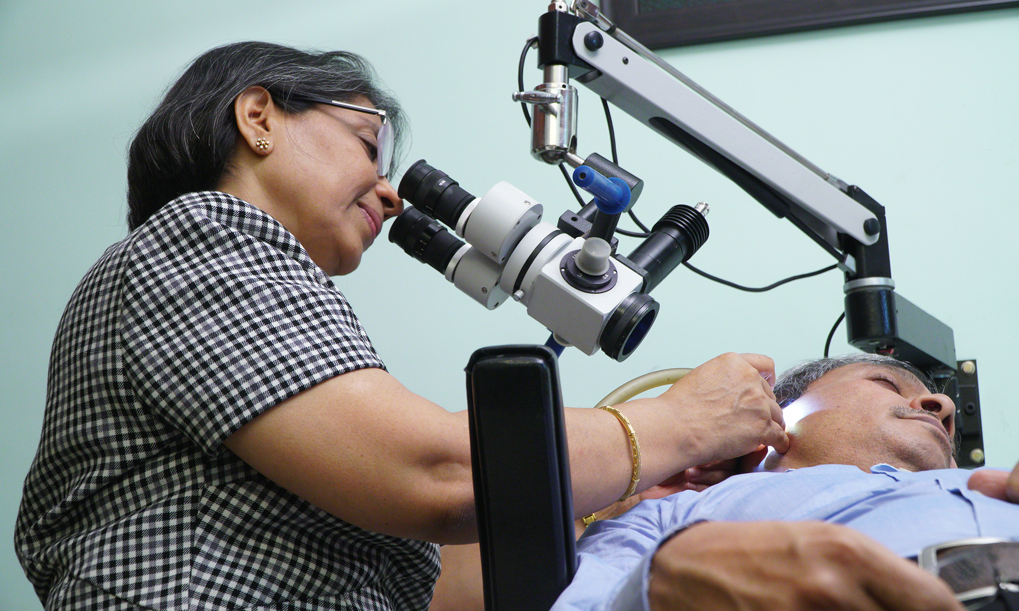
Recently, I was requested by a neurologist to examine a middle-aged lady admitted in the intensive care unit with meningitis as an alert caregiver had noticed some wetness in the right ear. The examination under the microscope revealed the extensive disease in the ear of which the meningitis was a complication. The mentally confused patient had not perceived anything amiss. Scans revealed extensive erosion of bone destroying the entire middle and inner ear on both the sides more so on the right. Further tests revealed the patient to be stone deaf in the right ear and partially deaf on the left. The insidious onset of the disease made the patient ignore her symptoms for long. Finally, the ear was operated upon to remove the involved bone, and the facial nerve hung like a ropeway at the end. The function of the nerve was almost normal after the surgery, but the hearing and imbalance remained uncorrected. Thankfully it became a safe ear now.
Judicious and prompt decisions about nasal diseases that affect the eustachian tube like the enlarged adenoids or nasal allergy are of utmost significance. Prevention is better than cure, and awareness about the disease is the first step to prevention. Most often, the condition is underdiagnosed, and sometimes the symptom is ignored even after the diagnosis due to naivete or negation. Surgery alone can cure the disease. This microscopic procedure involves the patient drilling of the affected bone to remove every bit of keratin while preserving significant structures like the facial nerve. Any remnant is sure to spark a flare-up later on. Even though treatment brings the disease under control, it cannot restore normalcy. The resultant cavity after the drilling is a lifelong liability to the patient as it will need to be cleared of debris by an otolaryngologist intermittently. The lack of awareness about this preventable condition is depressing to me as a doctor and to the patient.
It was an awesome read,ear looks so simple from outside but the article highlights the complexity and enlightens us on the workings of the ear
Thank you, Nikhil. I am happy that I could convey my message.
Really descriptive,ear looks and feels so simple ,but this article has enlightened me about the complexities and subtleness of the inner workings of ear.
Thank you, Dr Nikhil, for going through the post thoroughly
MADAM
I liked the title.
Also the way you have explained in detail about Cholesteatoma.
To read the line “Facial nerve hanging like a rope” sent shivers down the spine.
Can a person get Facial nerve palsy after this surgery?
If so what is the recovery rate?
Thanks, Dr Maggie. Facial nerve palsy is a complication of the disease, as well as, of the surgery. The recovery rate depends on the extent of damage to the nerve. If the damage is of a low-grade, good recovery can be expected with facial exercises.
Well articulated……could understand the problem and the implications.
Thank you Suchi, I feel great that I managed to convey the message.
Comprehensive as usual !
Thankyou, Shobha